The Fourteen Steps That Doctors Follow During A Supracondylar Amputation
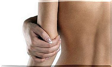
Supracondylar amputation is a surgical procedure in which a pelvic bone is cut over the joint head. Between 50% and 65% of these types of nontraumatic amputations are due to a complication due to diabetes.
A supracondylar amputation is performed when previous treatments to solve the problem have failed. This requires a process of acceptance and communication of a clear goal for the patient: Protecting their health and ensuring the best possible quality of life.
Based on the above, the target is therefore a healed, stable stump that a prosthesis can be put on as quickly as possible. The ultimate goal is for the patient to return to his normal life as soon as possible.
General principles
In general, amputations are classified as major and minor. Supracondylar amputations are larger since they cover a larger area. Regardless of their classification, however , all amputations are complex procedures that must conform to the basic principles. This is:
- They always involve treatment with antibiotics to control previous infections or as a preventative measure.
- Hemostasis, or the process of stopping bleeding, must be very strict. If bruising occurs, it is a sign of necrosis or infection.
- There should be no tension in the entrances on the skin edges. To prevent this from happening, doctors will need to handle the soft tissues carefully.
- There should be a reasonable relationship between bone and skin and muscle-tendon length. This prevents tension and provides good coverage of the bones.
- It is important to perform a pull of the nerves to prevent possible nerve tumors in the scar.
- The same must be done with the articular cartilage and tendons.
- Prevention of bone splinters in the wound or sharp edges.
- Repeat washing of the wound with a saline solution or cleanser before closing it.
Indications and contraindications
Physicians perform a supracondylar amputation when a previous infracondylar amputation failed to heal. It is also done when there is a contracture in the calf muscle, including a flexion of the joint in the knee.
The joint in the knee is lost in a supracondylar amputation. To avoid complications with the prosthesis that the patient will use, it is important that the stump is of sufficient length.
This procedure is not recommended for patients with gangrene or a thigh infection.
Technique for supracondylar amputation
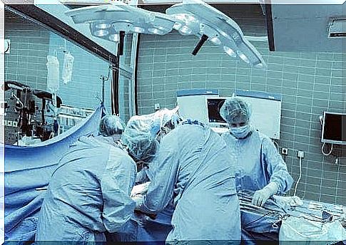
The steps that physicians follow when performing a supracondylar amputation are:
- First, they place the patient in a supine position.
- Then, they mark the cut.
- After that, they cut the skin with a cold scalpel.
- Doctors cut the subcutaneous tissue up to the aponeurosis or the tissue that covers the muscles and attaches them to the bone, with an electric scalpel. While cutting, doctors should leave enough tissue for the stump.
- They then identify, isolate and excise the superficial artery and the deep artery in the thigh, as well as the sciatic nerve. It is necessary for them to apply anesthesia.
- They cover the entire femur and reach around its entire circumference.
- After that, doctors separate the tissues that are stuck to the femur. To do so, they will need to use a periosteal elevator.
The last steps in a supracondylar amputation
- Physicians should customize a Percy amputation retractor to be able to amputate the femur. That way, they will ensure that there is enough soft tissue to cover the bone stump.
- After that, doctors split the bone with a Gigli saw. They do this at a 90 degree angle between the two ends of the saw. While doing so, they should constantly wash the area with a saline solution.
- Then, they file the edges of the bone.
- After that, they apply bone wax on the cut parts. They press and fix the bone wax on the cut parts. The leftovers should be discarded.
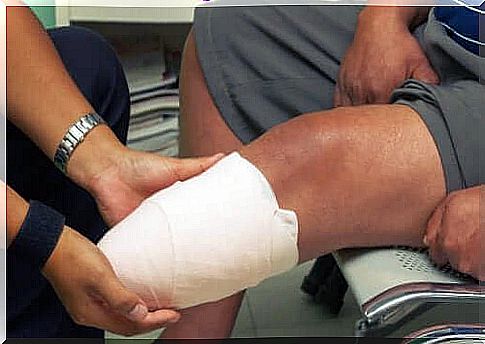
- After that, doctors close the stump with non-absorbable, sterile surgical suture, known as Prolene. First, they close the deeper muscle groups in order to cover the surface of the bone. Then, they close the superficial aponeurosis.
- Finally, doctors close the skin with a silk suture using vertical stitches.